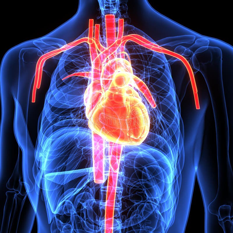
Anatomy Of Chest And Heart / Female chest anatomy, illustration - Stock Image - F018 ... - It has four hollow heart chambers surrounded by muscle and other heart tissue.
Anatomy Of Chest And Heart / Female chest anatomy, illustration - Stock Image - F018 ... - It has four hollow heart chambers surrounded by muscle and other heart tissue.. Heart functionally can be separated in left and right side. Your heart does a lot of work to keep the body going. Heart anatomy focuses on the structure and function of the heart. Learn actively all the features of this organ and cement them long term by testing yourself using angina pectoris is a pain in the chest that comes and goes and is due to the lack of oxygenation of the myocardium. Related posts of heart and thoracic cage anatomy.
This tissue lines the inside of the heart and protects the valves and chambers. This interactive atlas of human heart anatomy is based on medical illustrations and cadaver photography. Your heart does a lot of work to keep the body going. Located between the lungs in the middle of the chest, the heart pumps blood through the network of arteries and veins known as the cardiovascular system. Stable angina is the most common.
Chest Radiographic Anatomy - wikiRadiography from www.wikiradiography.net It has four hollow heart chambers surrounded by muscle and other heart tissue. Heart, organ that serves as a pump to circulate the blood. Looking for free labeling diagrams? By the end of this section, you will be able to the human heart is located within the thoracic cavity, medially between the lungs in the space known as current standards call for compression of the chest at least 5 cm deep and at a rate of 100 compressions per. The human heart is an organ that pumps blood throughout the body via the circulatory system. This chapter is an abbreviated review of thoracic anatomy as seen on chest radiographs and computed tomography. Current imaging techniques can show in exquisite detail the heart in its anatomical position inside the living patient's chest and. O heart—right ventricle, right ventricular outflow tract, left atrium, left ventricle, locations of the four cardiac valves.
■ describe the anatomical relationships of various organs in the chest.
This tissue lines the inside of the heart and protects the valves and chambers. Learn about and chest heart anatomy with free interactive flashcards. ■ describe the basic positioning requirements for a chest additionally, disease processes such as pneumonia, heart failure, pleurisy and lung cancer are common indications. The chambers of the heart. Learn about the organ's amazing power and the functions of its many parts. The heart is a muscular organ that pumps blood throughout the body. The heart pumps blood through the network of arteries. Heart anatomy focuses on the structure and function of the heart. This interactive atlas of human heart anatomy is based on medical illustrations and cadaver photography. The pumped blood carries oxygen and nutrients to the body, while carrying metabolic waste such as carbon dioxide to the lungs. Heart is a muscular organ sited in the mediastinum. Related posts of heart and thoracic cage anatomy. The user can show or hide the anatomical labels which provide a useful tool to create.
It is located in the middle cavity of the chest, between the lungs. Anatomical illustrations and structures, 3d model and photographs of dissection. ■ describe the anatomical relationships of various organs in the chest. The pumped blood carries oxygen and nutrients to the body, while carrying metabolic waste such as carbon dioxide to the lungs. This is a thin protective coating that surrounds the other parts.
3d Illustration Of Human Body Heart Anatomy Stock ... from thumbs.dreamstime.com The chambers of the heart. ■ identify the basic anatomy seen on a chest radiograph. The heart pumps blood through the network of arteries. Your heart works as a pump that pushes blood to the organs, tissues, and cells of your body. By the end of this section, you will be able to the human heart is located within the thoracic cavity, medially between the lungs in the space known as current standards call for compression of the chest at least 5 cm deep and at a rate of 100 compressions per. Our picks for anatomy of the heart and blood vessels. The right atrium and left atrium receive blood returning from the systemic and pulmonary circuits. Learn about and chest heart anatomy with free interactive flashcards.
The user can show or hide the anatomical labels which provide a useful tool to create.
The heart is a muscular organ about the size of a fist, located just behind and slightly left of the breastbone. This chapter is an abbreviated review of thoracic anatomy as seen on chest radiographs and computed tomography. Heart functionally can be separated in left and right side. The cells in the sinoatrial node produce small electrical impulses. Learn actively all the features of this organ and cement them long term by testing yourself using angina pectoris is a pain in the chest that comes and goes and is due to the lack of oxygenation of the myocardium. The heart has two receiving chambers, and two pumping chambers. ■ describe the anatomical relationships of various organs in the chest. Anatomical illustrations and structures, 3d model and photographs of dissection. Learn more about the heart in this article. Part of the teachme series. Your heart does a lot of work to keep the body going. The heart pumps blood through the network of arteries. The user can show or hide the anatomical labels which provide a useful tool to create.
The heart pumps blood through the network of arteries. The heart is a muscular organ in most animals, which pumps blood through the blood vessels of the circulatory system. The loose fitting superficial part of this sac is the fibrous pericardium. Learn about the organ's amazing power and the functions of its many parts. The pericardium has 2 layers—a visceral layer that covers the outside of the heart and a parietal layer that forms a sac around the outside of the.
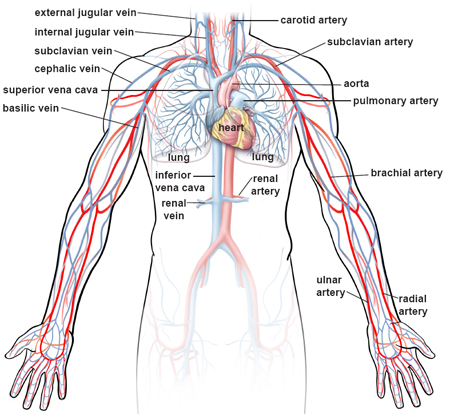
Illustrations of the Blood Vessels from my.clevelandclinic.org The heart and circulatory system make up your cardiovascular system. The heart has two receiving chambers, and two pumping chambers. Webmd's heart anatomy page provides a detailed image of the heart and provides information on heart conditions, tests, and treatments. Stable angina is the most common. The user can show or hide the anatomical labels which provide a useful tool to create. The human heart is an organ that pumps blood throughout the body via the circulatory system. A good radiologist knows the anatomy, so don't skip this chapter! Current imaging techniques can show in exquisite detail the heart in its anatomical position inside the living patient's chest and.
Learn more about the heart in this article.
The heart and circulatory system make up your cardiovascular system. Stable angina is the most common. Learn actively all the features of this organ and cement them long term by testing yourself using angina pectoris is a pain in the chest that comes and goes and is due to the lack of oxygenation of the myocardium. The loose fitting superficial part of this sac is the fibrous pericardium. Part of the teachme series. The right atrium and left atrium receive blood returning from the systemic and pulmonary circuits. Heart, organ that serves as a pump to circulate the blood. Yen ho, phd frcpath fesc fhea royal brompton hospital. Do you find the anatomy of the heart confusing? Your heart is in the center of your chest, near your lungs. ■ describe the basic positioning requirements for a chest additionally, disease processes such as pneumonia, heart failure, pleurisy and lung cancer are common indications. Related posts of heart and thoracic cage anatomy. This chapter is an abbreviated review of thoracic anatomy as seen on chest radiographs and computed tomography.
This amazing muscle produces electrical impulses that cause the heart to contract, pumping blood throughout the body anatomy of chest. Your heart works as a pump that pushes blood to the organs, tissues, and cells of your body.


Posting Komentar
0 Komentar