Anatomy Mapping Foramina : List Of Foramina Of The Human Body Wikiwand / This video provides a walkthrough of the foramen of the skull (cranial foramina), including the cranial nerves that pass through each foramen.
Anatomy Mapping Foramina : List Of Foramina Of The Human Body Wikiwand / This video provides a walkthrough of the foramen of the skull (cranial foramina), including the cranial nerves that pass through each foramen.. In this course, craig elliot, provides a breakdown of the female anatomy. Microsurgical anatomy applied to tumors of the medial temporal lobe 11. A classic painting/stained glass window of foramen spinosum: Post ethmoidal foramen ant ethmoidal foramen frontoethmoidal suture anterior lacrimal crest optic foramen. For mnemonics in other medical specialties, see this list of medical mnemonics.
Learn vocabulary, terms and more with flashcards, games and other study tools. The journal of laryngology clinical anatomy of blockade of the pterygopalatine ganglion: Home » anatomy monday » anatomy monday: Microsurgical anatomy applied to tumors of the medial temporal lobe 11. 403 просмотров · 26 декабря 2019 г.
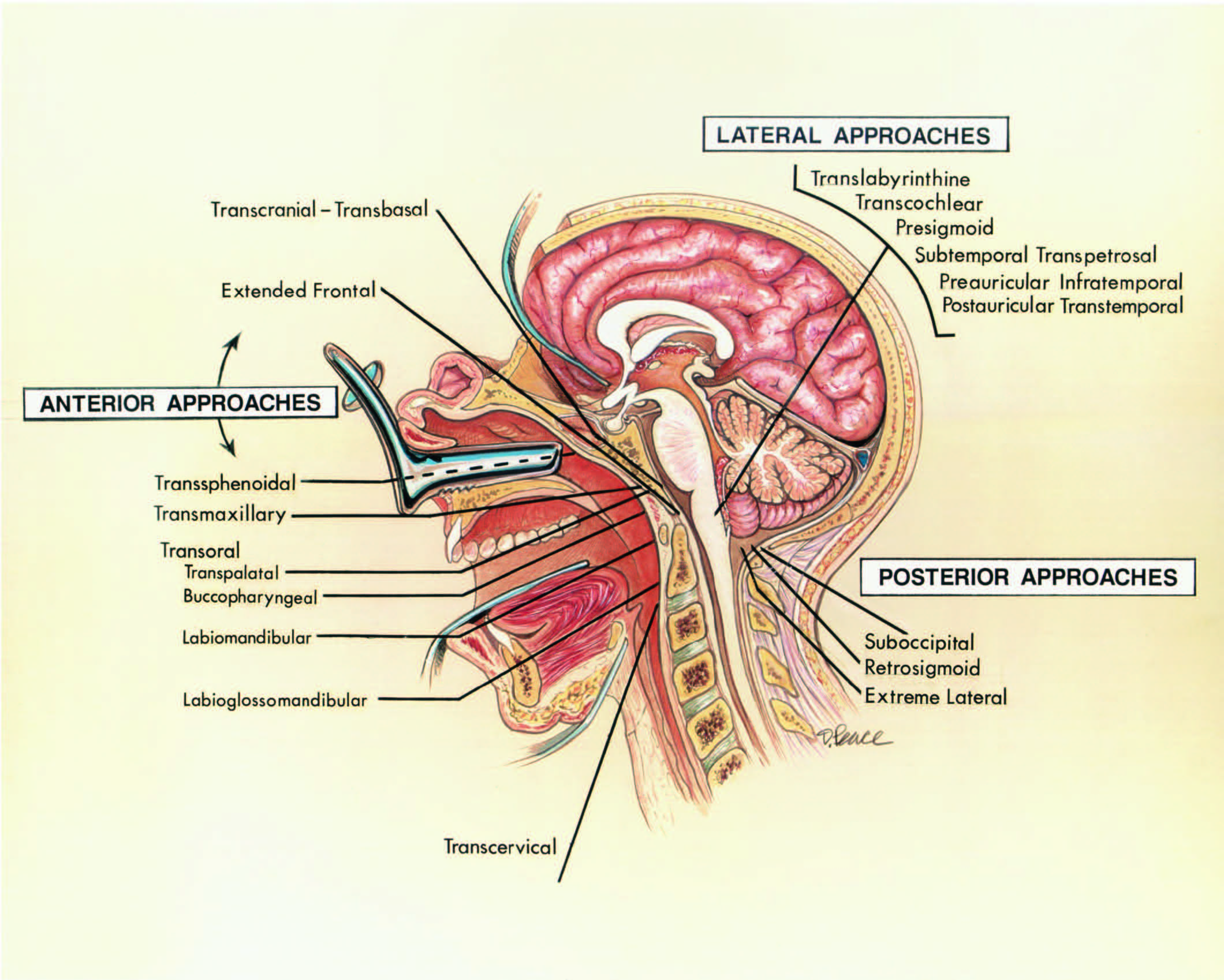
Diagnosis Of Skull Base Osteomyelitis Radiographics from pubs.rsna.org Applied knowledge of anatomical relations regarding surgical procedures and approaches was discussed and their relations were demonstrated via cadaveric dissections. In this course, craig elliot, provides a breakdown of the female anatomy. …transverse processes of the lower sacral vertebrae, on each side, are a series of four openings (sacral foramina). For mnemonics in other medical specialties, see this list of medical mnemonics. In this article we will discuss the anatomy, its contents and clinical relevance of the mandibular mandibular foramen. Inferior vena cava posteriorly,hepatic artery & portal vein anteriorly, quadrate lobe superiorly. Each fossa contains specific foramina, through which various anatomical structures pass through. 403 просмотров · 26 декабря 2019 г.
…transverse processes of the lower sacral vertebrae, on each side, are a series of four openings (sacral foramina).
…transverse processes of the lower sacral vertebrae, on each side, are a series of four openings (sacral foramina). Endoscopic approach with bony landmarks. Uruj zehra mbbs, mphil, phd last. A classic painting/stained glass window of foramen spinosum: Inferior vena cava posteriorly,hepatic artery & portal vein anteriorly, quadrate lobe superiorly. Literature review and pictorial tour. Omental foramen or epiploic foramen or foramen of winslow or additus to lesser sac 【at t5 level】 (note: Thoracic nerve roots emerge from the intervertebral foramina into the paravertebral space. Each fossa contains specific foramina, through which various anatomical structures pass through. Location on base of skull foramen spinosum is adjacent to the spine of sphenoid. In the brain, the interventricular foramina (or foramina of monro) are channels that connect the paired lateral ventricles with the third ventricle at the midline of the brain. 3d video anatomy tutorial on the foramina of the skull and the cranial nerves and blood vessels that in this tutorial, i'm going to be looking at the different structures that pass through the foramina in the. Home » anatomy monday » anatomy monday:
Another pathology of the posterior fossa can occur when the cerebellar tonsils descend below the foramen magnum; In this article we will discuss the anatomy, its contents and clinical relevance of the mandibular mandibular foramen. The map data is available for public download. For mnemonics in other medical specialties, see this list of medical mnemonics. Frank pameijer, erik beek, frank joosten and robin smithuis.
2 from Location on base of skull foramen spinosum is adjacent to the spine of sphenoid. Thoracic nerve roots emerge from the intervertebral foramina into the paravertebral space. The map data is available for public download. A classic painting/stained glass window of foramen spinosum: As channels, they allow cerebrospinal fluid (csf). In this course, craig elliot, provides a breakdown of the female anatomy. Omental foramen or epiploic foramen or foramen of winslow or additus to lesser sac 【at t5 level】 (note: In this article we will discuss the anatomy, its contents and clinical relevance of the mandibular mandibular foramen.
The interventricular foramen, also known as foramen of monro, is part of the ventricular system and the connection between the third ventricle and the lateral ventricle.
Home » anatomy monday » anatomy monday: Thoracic nerve roots emerge from the intervertebral foramina into the paravertebral space. Last week i showed the superior foramina of the nasopalatine canal and this week is the inferior foramen; This video provides a walkthrough of the foramen of the skull (cranial foramina), including the cranial nerves that pass through each foramen. Microsurgical anatomy applied to tumors of the medial temporal lobe 11. Other articles where sacral foramen is discussed: Another pathology of the posterior fossa can occur when the cerebellar tonsils descend below the foramen magnum; The map data is available for public download. As channels, they allow cerebrospinal fluid (csf). Applied knowledge of anatomical relations regarding surgical procedures and approaches was discussed and their relations were demonstrated via cadaveric dissections. …transverse processes of the lower sacral vertebrae, on each side, are a series of four openings (sacral foramina). The interventricular foramen, also known as foramen of monro, is part of the ventricular system and the connection between the third ventricle and the lateral ventricle. Frank pameijer, erik beek, frank joosten and robin smithuis.
Each fossa contains specific foramina, through which various anatomical structures pass through. Endoscopic approach with bony landmarks. Omental foramen or epiploic foramen or foramen of winslow or additus to lesser sac 【at t5 level】 (note: As channels, they allow cerebrospinal fluid (csf). This is termed a chiari i.
Foramen Magnum The Neurosurgical Atlas from assets.neurosurgicalatlas.com Home » anatomy monday » anatomy monday: Last week i showed the superior foramina of the nasopalatine canal and this week is the inferior foramen; Each fossa contains specific foramina, through which various anatomical structures pass through. Literature review and pictorial tour. Other articles where sacral foramen is discussed: This video provides a walkthrough of the foramen of the skull (cranial foramina), including the cranial nerves that pass through each foramen. The lesion originates at the left neural foramina and grows along the course of the brachial plexus (red arrow). The interventricular foramen, also known as foramen of monro, is part of the ventricular system and the connection between the third ventricle and the lateral ventricle.
Post ethmoidal foramen ant ethmoidal foramen frontoethmoidal suture anterior lacrimal crest optic foramen.
Other articles where sacral foramen is discussed: A classic painting/stained glass window of foramen spinosum: Thoracic nerve roots emerge from the intervertebral foramina into the paravertebral space. Applied knowledge of anatomical relations regarding surgical procedures and approaches was discussed and their relations were demonstrated via cadaveric dissections. Another pathology of the posterior fossa can occur when the cerebellar tonsils descend below the foramen magnum; 3d video anatomy tutorial on the foramina of the skull and the cranial nerves and blood vessels that in this tutorial, i'm going to be looking at the different structures that pass through the foramina in the. This article discusses each of the aforementioned fossae and their associated foramina. Uruj zehra mbbs, mphil, phd last. Each fossa contains specific foramina, through which various anatomical structures pass through. 403 просмотров · 26 декабря 2019 г. Here they send white rami communicantes to the sympathetic chain. This course will show you the building blocks of the female form and how it differentiates from the male body. For mnemonics in other medical specialties, see this list of medical mnemonics.
Applied knowledge of anatomical relations regarding surgical procedures and approaches was discussed and their relations were demonstrated via cadaveric dissections anatomy map. As channels, they allow cerebrospinal fluid (csf).


Posting Komentar
0 Komentar